The Clinique Dentaire des Vallées de l’Outaouais has made a major investment in state-of-the-art equipment, as well as in the training of its staff to provide its patients with the ultimate in the art of dentistry. Thanks to their
High quality sterilization
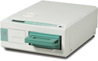
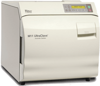
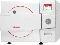
We hold our the aseptic and sterilization procedures to meticulous standards. We have two ultrasonic baths, a Statim 2000 and a Statim 5000 as well as two autoclaves: a Midmark 11 and a Tattnauer.
Digital Radiography
We use digital radiology to reduce your exposure to X-rays, improve diagnostic quality and as a demonstration tool.
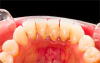
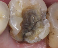
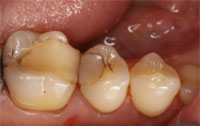
Intra Oral Camera
It is used to take images inside your mouth to allow you to visualize its condition and then participate in your treatment plan.
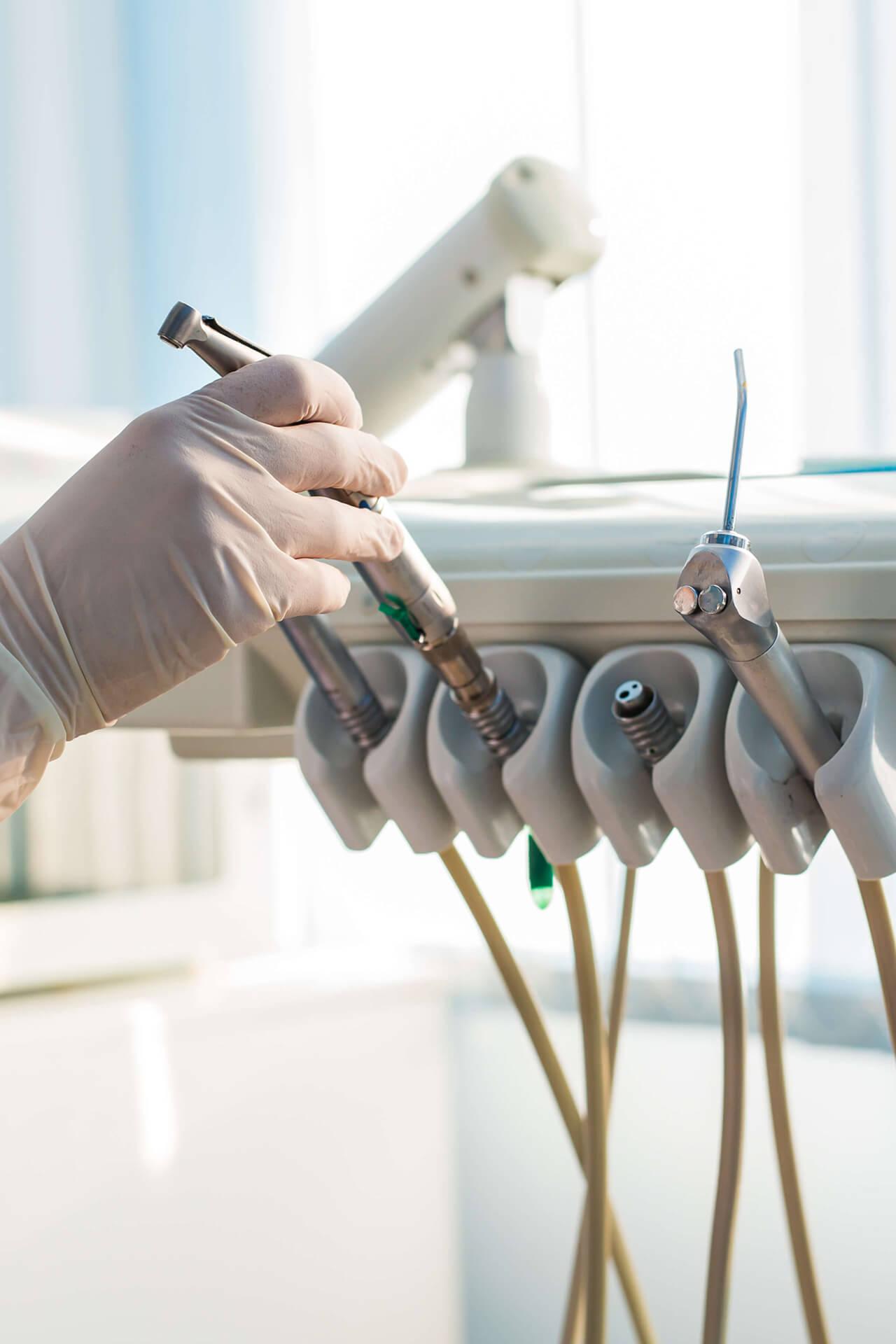
Our equipment
For the well-being of our clients, all the staffs of our clinic respect meticulously the strict measures of asepsis and sterilization of the Ordre des Dentistes du Québec (ODQ).
The sterilization of our medical instruments involves the following steps
- Cleaning instruments by brushing mechanically under water and soaking in ultrasonic baths for 15 minutes.
- Once disinfected, the instruments are rinsed, dried and packaged in cassettes or disposable paper / plastic bags.
- The instruments are then loaded into the various sterilizers we have (Statim 2000, Statim 5000, Midmark II, Tattnaeur) for cycles varying from 12 to 55 minutes and temperatures varying from 135°C/375°F to 190°C/375°F, depending on the cycle and the unit.
- Finally, cassettes and sterile instrument bags are stored in our clean, closed shelves until they are used in the operating rooms.
Verification of sterilization is done on each cycle using adhesive tapes on the cassettes and ink-marked bags that change color when exposed and penetrated by the appropriate temperature.
We also hire an independent microbiological laboratory to analyze the effectiveness of our devices at least once a month.
Digital radio also provides an overview of the dental structure.
Here are all the advantages of digital radio:
- 90% reduction in the exposure rate compared to traditional radiographs;
- Improving the quality of our diagnoses for your oral health;
- Instant result, therefore immediate evaluation of the radio and less waiting for you;
- Demonstration tool and explanation by showing you the radiographs of your teeth on a 22-inch screen;
- Reduced deadlines for direct emails to insurance companies and specialists.
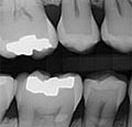
The interproximal radiography
It allows simultaneous observation of the crowns of the upper and lower teeth.
Interproximal radiography is essential for dental examination to identify caries in the interdental areas and the joints of filling that are otherwise undetectable.
These radiographs are usually taken annually.
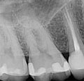
The periapical radiography
It allows to isolate a group of upper or lower teeth so as to be able to study their roots, their bone supports, but also the lesions related to the nerve of the tooth such as an abscess.
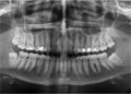
The panoramic radiography
It is an essential complement to dentistry.
The panoramic radiography gives an overview of the upper and lower teeth, the maxilla, the articulations and the sinuses.
It allows us to know the location of the teeth for children with mixed dentition and also the position of the teeth of wisdom to plan their extraction if necessary.
In addition, it allows evaluation of bone loss, root abnormalities, cysts, tumors and abscesses.
High-quality Imagery
The camera is connected to the dentist’s computer and those images are projected onto our 22-inch screens to facilitate diagnosis and to show you what the dentist sees in your mouth. This allows you to visualize the health of your teeth and gums, allowing you to better understand the usefulness of the treatment.
Electronic Record
The images of the intraoral camera are stored in your electronic record and allow you to follow the evolution of your oral condition (recession of gums, evaluation of photos before and after).